Entropion and Ectropion Surgery for Dogs: What Fort Worth Pet Owners Should Know
When Your Dog’s Eyes Always Seem to Be Bothering Them
You’ve noticed it for a while now. One eye that always looks a little watery. A dog who squints in normal lighting, rubs their face on the couch cushions, or blinks more than seems right. It’s easy to chalk this up to allergies or a piece of debris that will resolve on its own. But if the symptoms keep coming back, something structural may be going on.
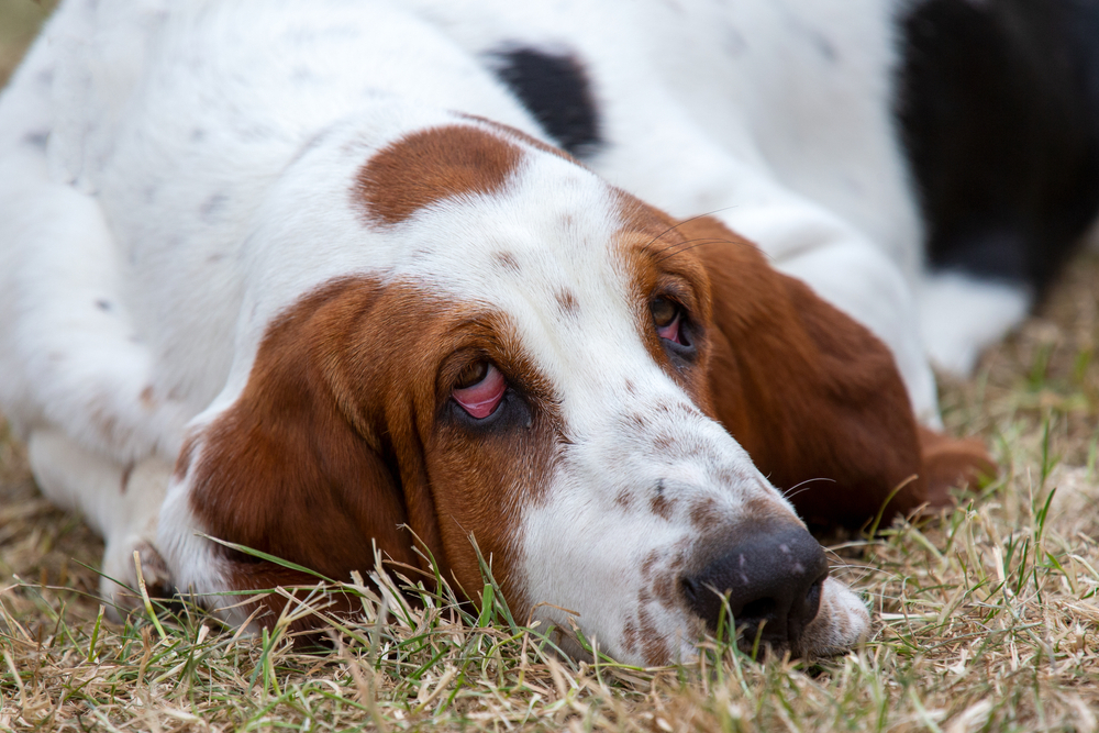
Entropion is a condition where the eyelid rolls inward, causing the skin and lashes to rub directly against the surface of the eye with every blink. Ectropion is the opposite problem: the lower eyelid droops outward, exposing the sensitive inner lid tissue to air, dust, and bacteria. Both conditions are among the most common eye conditions seen in veterinary practice, and both are highly treatable with the right approach.
At Woodland Springs Veterinary Hospital in Fort Worth, our surgical services include eyelid repair for both dogs and cats. Because we’re AAHA accredited, you can rest assured knowing that we use nothing but the best in modern, safe anesthesia and medical protocols, including laser-assisted surgery. Contact us to schedule an evaluation if your pet’s eyes have been a recurring concern.
What Are Entropion and Ectropion, and Why Do They Happen?
The Eye Structures Involved
The eyelids do a lot more than blink. They distribute tear film across the cornea, protect the eye surface from debris, and create the seal the eye needs to stay healthy. When lid position is off, even slightly, the consequences build up over time.
With entropion, the rolled-in lid means that with every single blink, fur and lashes scrape across the cornea. The cornea is extraordinarily sensitive tissue, and that friction is genuinely painful, producing tearing, redness, squinting, and over time, real structural damage. With ectropion, the drooping lid leaves the pink conjunctival tissue exposed and drying out. Dust, pollen, and bacteria settle into that exposed pocket, leading to chronic irritation and repeated infections that antibiotics can treat temporarily but not solve.
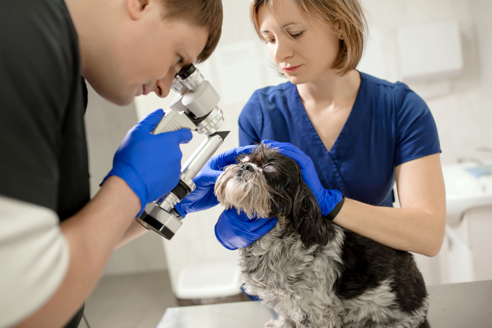
Both conditions can be present at birth or develop over a pet’s lifetime. Accurate diagnosis matters because several eye issues can look similar from the outside, and a proper evaluation examines lid position, corneal health, and tear production together rather than treating symptoms in isolation. Our diagnostics include thorough ophthalmic evaluations before any surgical planning begins.
Which Dogs (and Cats) Are Most at Risk?
Genetics and facial structure account for the vast majority of eyelid abnormality cases. Eyelid disorders are far more prevalent in certain breeds because their physical conformation includes the anatomical features that cause lid problems. Hereditary eyelid conditions run in breed lines, which is why these issues concentrate so predictably in specific populations.
Breeds commonly affected by entropion (inward rolling):
- Shar Peis, where heavy facial folds create significant tension on the eyelids
- Chow Chows, Rottweilers, English Bulldogs, and Mastiffs
- Great Danes and Saint Bernards
- Brachycephalic dogs, like Pugs, Frenchies, and Bulldogs and flat-faced cat breeds like Persians and Himalayans, whose compressed facial anatomy affects lid position
Breeds commonly affected by ectropion (outward drooping):
- Bloodhounds and Basset Hounds, whose loose skin produces the characteristic low-hanging lids
- Cocker Spaniels, Newfoundlands, Saint Bernards, and Great Danes
Some dogs develop both conditions in different parts of the same eyelid simultaneously, a configuration sometimes called “diamond eye” due to the shape it creates. Brachycephalic pets often develop “medial canthal entropion”- meaning the inner corner of their eyelid rolls in. We can assess and fix this as a part of our brachycephalic airway surgery services, when also fixing their stenotic nares or elongated soft palates with our surgical laser.
Beyond breed, contributing factors include age-related tissue loosening, chronic inflammation that shifts lid position over time, previous eye injuries, and even intense squinting from pain, which can temporarily pull a lid inward and mimic structural entropion.
If you have a puppy from a breed on this list, breed-specific eye concerns are worth raising at your first general wellness visit so we can monitor as the face matures.
What Signs Should Prompt a Veterinary Evaluation?
A pet communicating eye discomfort does so in fairly predictable ways. The signs of eye pain in dogs and cats worth watching for include:
- Excessive tearing that leaves chronic wet staining below the eye
- Squinting, keeping one eye partially closed, or blinking asymmetrically
- Pawing or rubbing at the face, particularly the eye area
- Visible redness of the white of the eye or the inner lid tissue
- Cloudiness, haze, or color changes in the normally clear cornea
- Recurrent eye infections, thick discharge, or crusting around the lid margins
- A visible rolling of the lid edge inward or outward
These are not cosmetic concerns. They are pain signals, and dismissing them as “just how this breed looks” is one of the most common reasons eyelid conditions progress to something more serious before they are addressed.
What Happens When Eyelid Problems Go Untreated?
The complications of untreated entropion and ectropion are progressive and not minor. For entropion, the ongoing friction of lid and lash tissue across the cornea leads to corneal ulcers, which are painful open sores on the eye surface. Superficial ulcers are manageable, but deep or infected ulcers carry a risk of perforation. Chronic irritation also deposits pigment on the cornea over time, permanently reducing clarity even after the structural problem is corrected.
For ectropion, the persistent eye discharge and exposed tissue create ideal conditions for bacterial infections that recur reliably until the underlying lid anatomy is addressed. The longer either condition is left unresolved, the more complicated the surgical approach becomes and the less completely any pre-existing damage can resolve. Contact us promptly if your pet is showing any of these signs.
How Do Veterinarians Diagnose Eyelid Abnormalities?
A thorough eye examination is more involved than a quick glance at whether a lid looks off. One of the most informative early steps is applying topical anesthetic drops to the eye before evaluating lid position. Many dogs squint so intensely from corneal pain that the squinting itself distorts the lid, making it appear abnormal when the true structural change is more subtle. Numbing drops relax that squinting and allow the resting lid position to be assessed accurately.
The complete evaluation also includes:
- Tear production measurement to rule out dry eye, which can mimic or worsen entropion symptoms
- Fluorescein staining of the corneal surface to identify any existing ulcers before surgery
- Magnified examination for eyelash problems like distichiasis, where abnormal lash positioning causes symptoms similar to entropion without actual lid rolling
- Assessment of overall facial conformation and breed-related anatomy
Standard ocular tests give the veterinary team objective measurements that inform both diagnosis and the specific surgical plan. Our diagnostics team conducts these evaluations carefully before any recommendations are made.
What Are the Treatment Options?
Temporary Tacking for Young or Growing Pets
Not every dog needs immediate permanent surgery. In puppies whose facial anatomy is still developing, attempting definitive correction too early risks overcorrecting once growth is complete and the final facial proportions are established. For these patients, temporary eyelid tacking is the appropriate first step: small sutures hold the lid in a more normal position to protect the cornea from friction while the face matures. Tacking is a minor procedure, can be repeated as needed, and buys time until permanent correction makes sense. Our team will walk you through whether staging is the right approach.
Permanent Surgical Repair
When the eyelid condition is clearly structural and the anatomy is stable, definitive surgery delivers lasting relief. The technique depends on which condition is present, how severe the malposition is, which part of the lid is affected, and the individual pet’s anatomy. Both entropion and ectropion are corrected by precisely removing or repositioning a carefully measured amount of tissue.
The technical demands of eyelid surgery are significant because the margin for error is narrow. Our preferred approach is conservative correction with the option to revisit if needed, rather than aggressive single-procedure correction that risks creating the opposite deformity. Anesthesia is individualized to each patient, with pre-anesthetic bloodwork, IV catheter placement, and continuous monitoring throughout.
Entropion in Cats
Feline eyelid disease follows a different pattern than in dogs. Entropion in cats tends to develop later in life rather than at birth, and it often occurs alongside other eye surface disease rather than as a standalone structural issue. Cats frequently require a combination of surgical techniques tailored to their specific anatomy and concurrent conditions. Cat owners with questions about their pet’s recurring eye problems are welcome to reach out directly.
What Should You Expect on Surgery Day?
Walking through the process ahead of time makes a real difference in how prepared owners feel. At Woodland Springs, each surgical day begins with a review of pre-anesthetic bloodwork and confirmation of the surgical plan with the owner. Anesthesia is customized to the individual patient’s age, weight, breed, and health status, with pain management built in before the procedure begins rather than added reactively afterward.
Throughout surgery, a dedicated team member continuously monitors heart rate, blood pressure, oxygen saturation, respiratory rate, and temperature. The eyelid area is carefully prepared, precise measurements are taken before any tissue is removed, and fine instruments and suture material are used throughout. Most patients go home the same day once fully alert and stable, with written post-operative instructions and all needed medications.
What Does Recovery Look Like at Home?
The First Few Days
Mild swelling and bruising around the eyelid are expected after surgery and typically peak around the second day before beginning to resolve. A small amount of clear or slightly pink discharge is normal. Most pets are quieter than usual while pain medication is active.
Call the clinic if you notice rapidly worsening swelling rather than stable or improving swelling, thick yellow or green discharge, bleeding at the incision site, or sutures that appear loose or out of place.
The E-collar is non-negotiable during recovery. One paw swipe can undo precise surgical work in a moment, and the cone is the only reliable protection for small, carefully placed eyelid sutures. Tips on administering eye medications to pets can make the post-operative drop and ointment routine easier to manage. Having a second person gently hold the pet, following each application with a high-value treat, and keeping sessions brief helps most animals adjust within a day or two.
Healing Timeline and Follow-Up
Sutures are typically removed at a recheck appointment around 10 to 14 days post-surgery. The lid position at that point is close to the final result, though residual swelling may still be present. Full settling of the corrected lid position is usually apparent by weeks three to six, once all swelling has resolved. Minor revisions are occasionally recommended after the initial healing is complete, and the conservative surgical approach makes those straightforward when needed.
What Outcomes Can You Expect?
Eyelid surgery has excellent outcomes when performed at the right time with attentive home recovery. For most pets, the improvement is apparent quickly: the squinting diminishes, the constant tearing settles, and the face-rubbing stops. That visible relief is one of the most rewarding outcomes in veterinary surgery, for the team and for the owner watching their dog finally feel comfortable.
Factors that influence the final result include the severity of the lid malposition before surgery, the degree of corneal changes already present, the pet’s age at the time of correction, and how consistently the cone and medications were used during recovery. Pre-existing corneal scarring may not completely resolve after surgery, but stopping the ongoing damage and relieving the pain are meaningful outcomes on their own. Owners of show dogs should note that some breed registries have rules about surgical corrections and show eligibility, worth reviewing before scheduling.
Our team monitors ongoing eye health at regular wellness visits for patients who have had eyelid corrections, and our wellness plans make consistent preventive care accessible.
Ready to Help Your Pet See Clearly and Comfortably
Entropion and ectropion are among the most consistently correctable conditions in veterinary surgery, and the difference in a pet’s quality of life before and after is often dramatic. If your dog or cat squints chronically, tears excessively, or rubs at their face regularly, a thorough evaluation is the right next step. These are not problems to wait out.
Woodland Springs Veterinary Hospital brings AAHA-accredited standards and genuine investment in your pet’s outcome to every case we see. Schedule an appointment online or contact our Fort Worth team.
Leave A Comment